.jpg)
|
|
|
|
||
|
|
|
|
|
|
|
|
|
|
|
|
|
|
|
|
||
|
|
|
|||
Incomplete Root Removal for Socket Shield
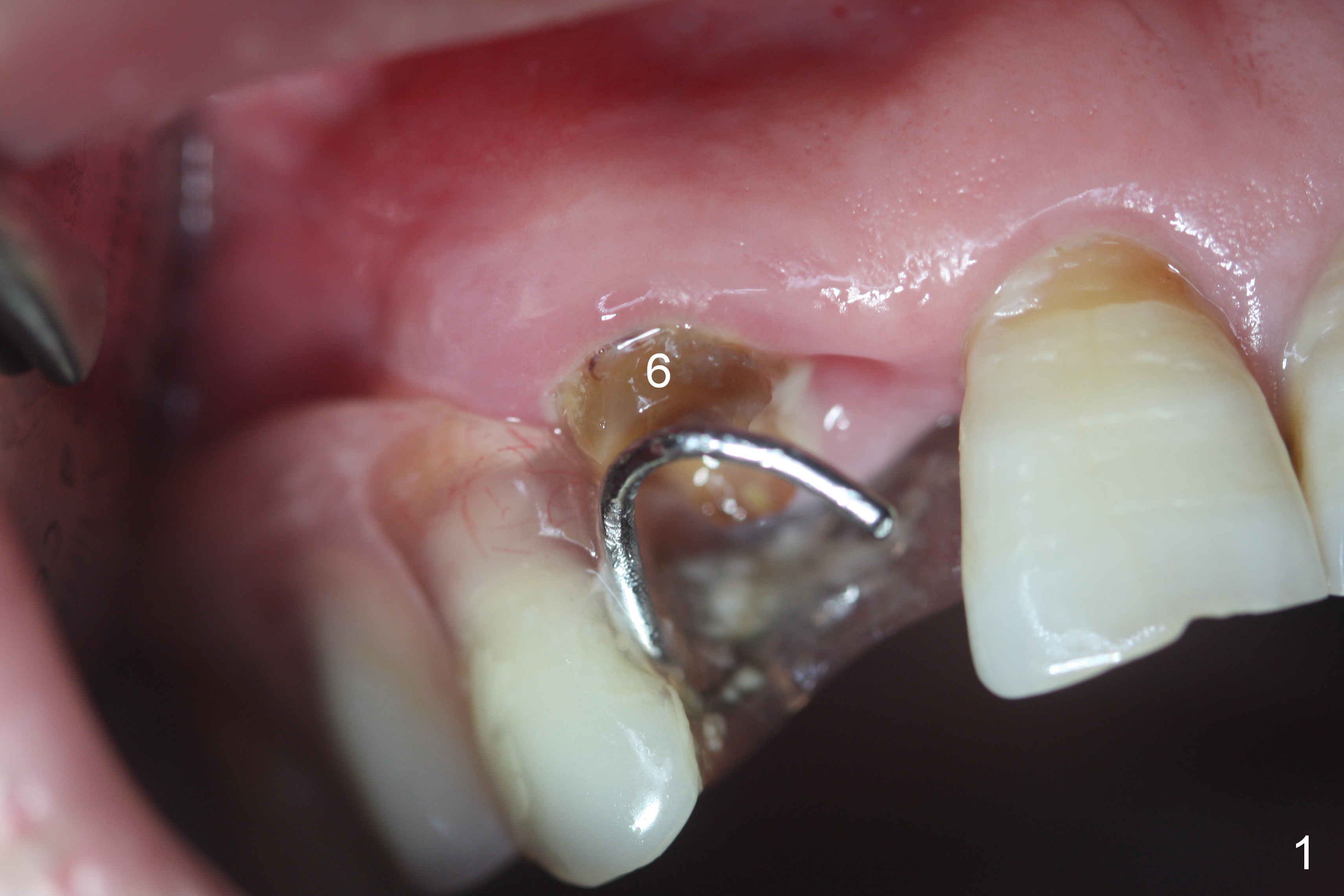
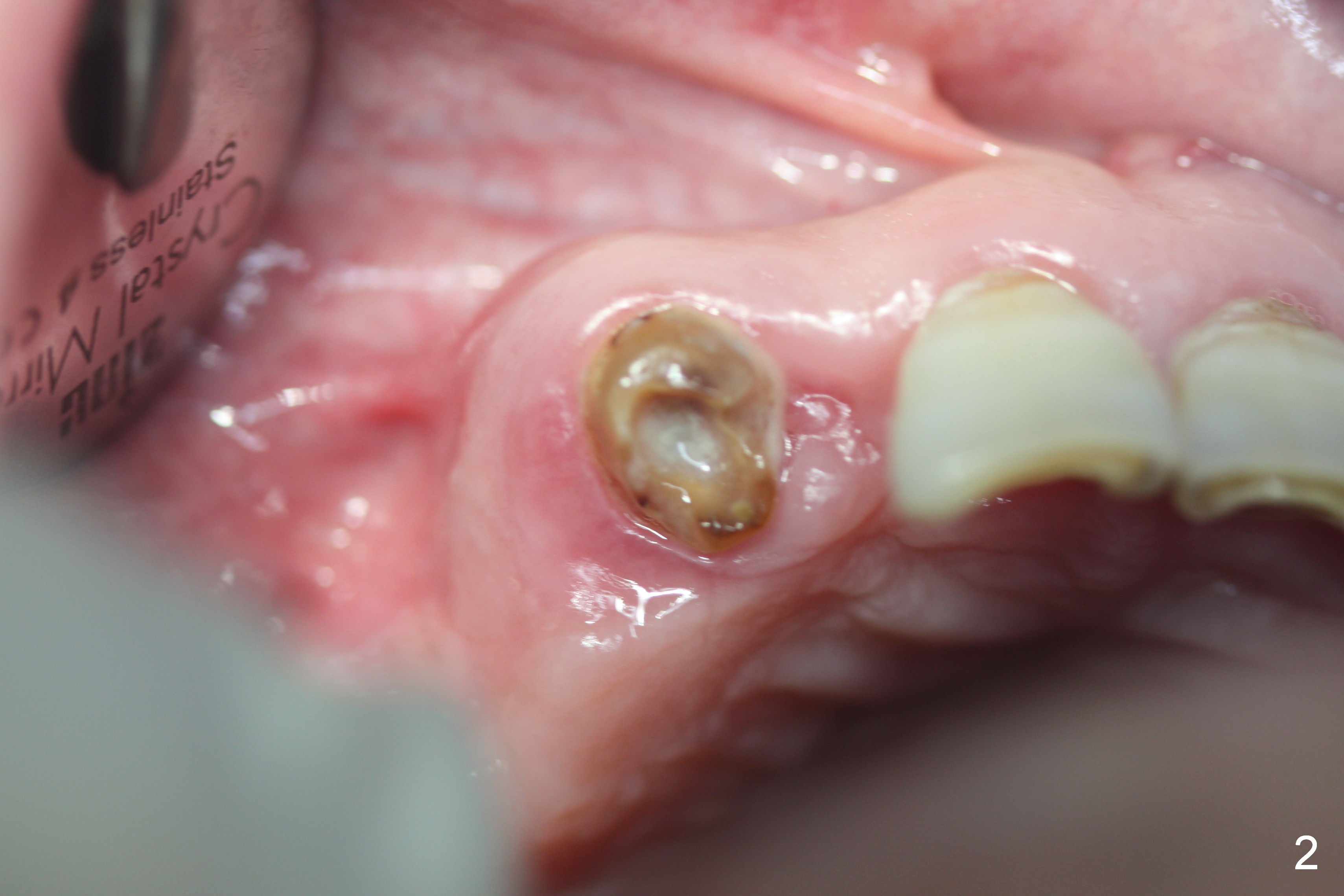
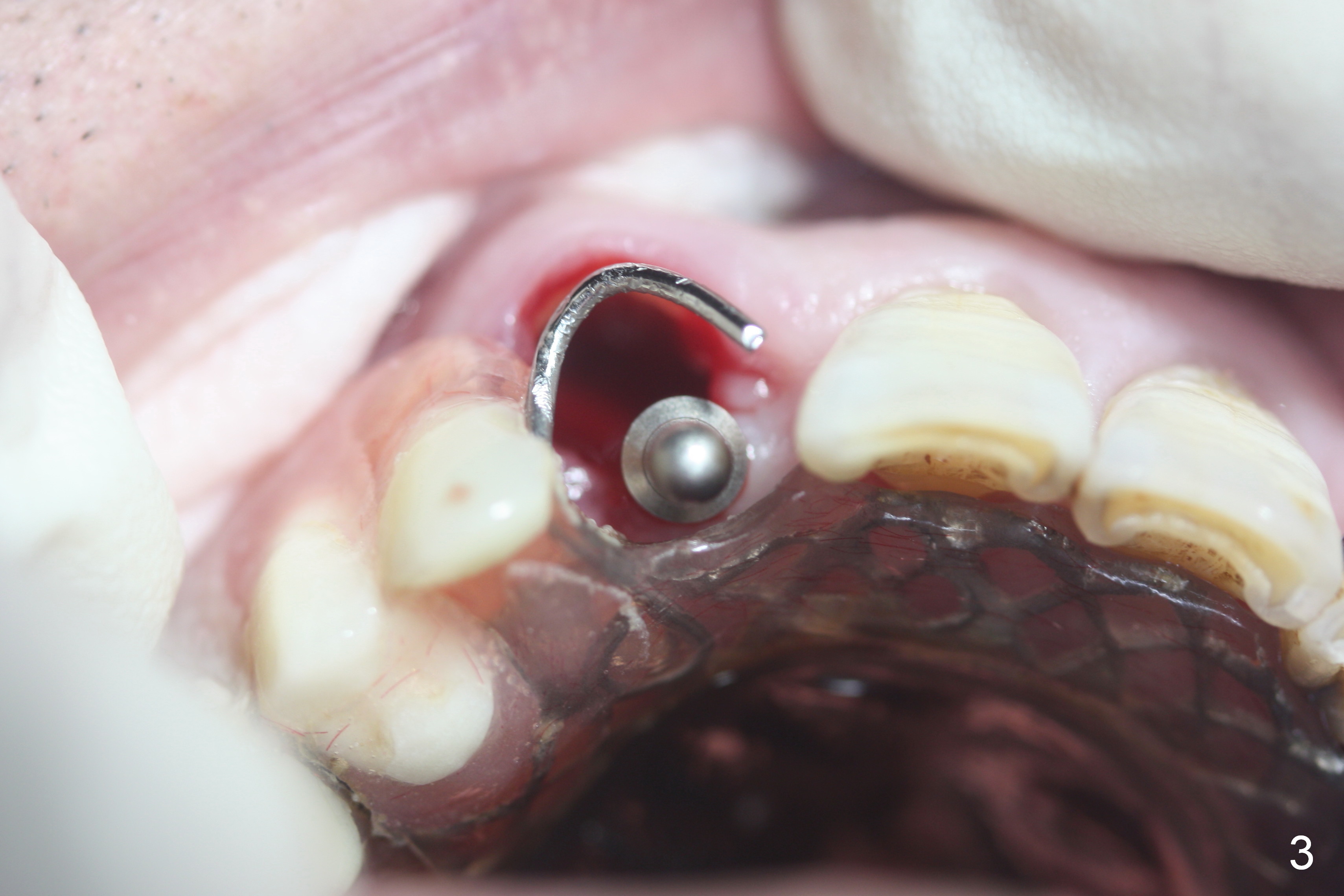
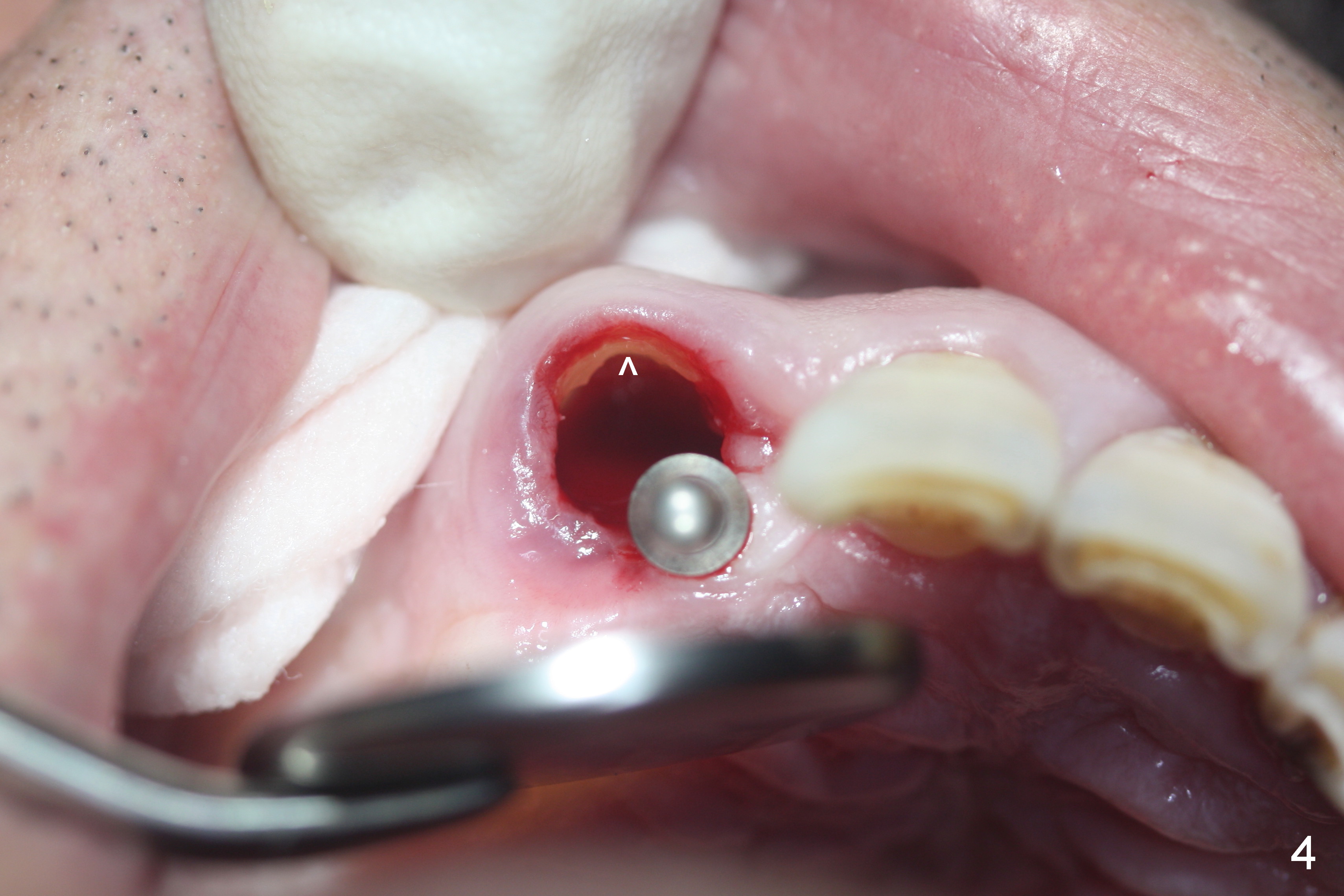
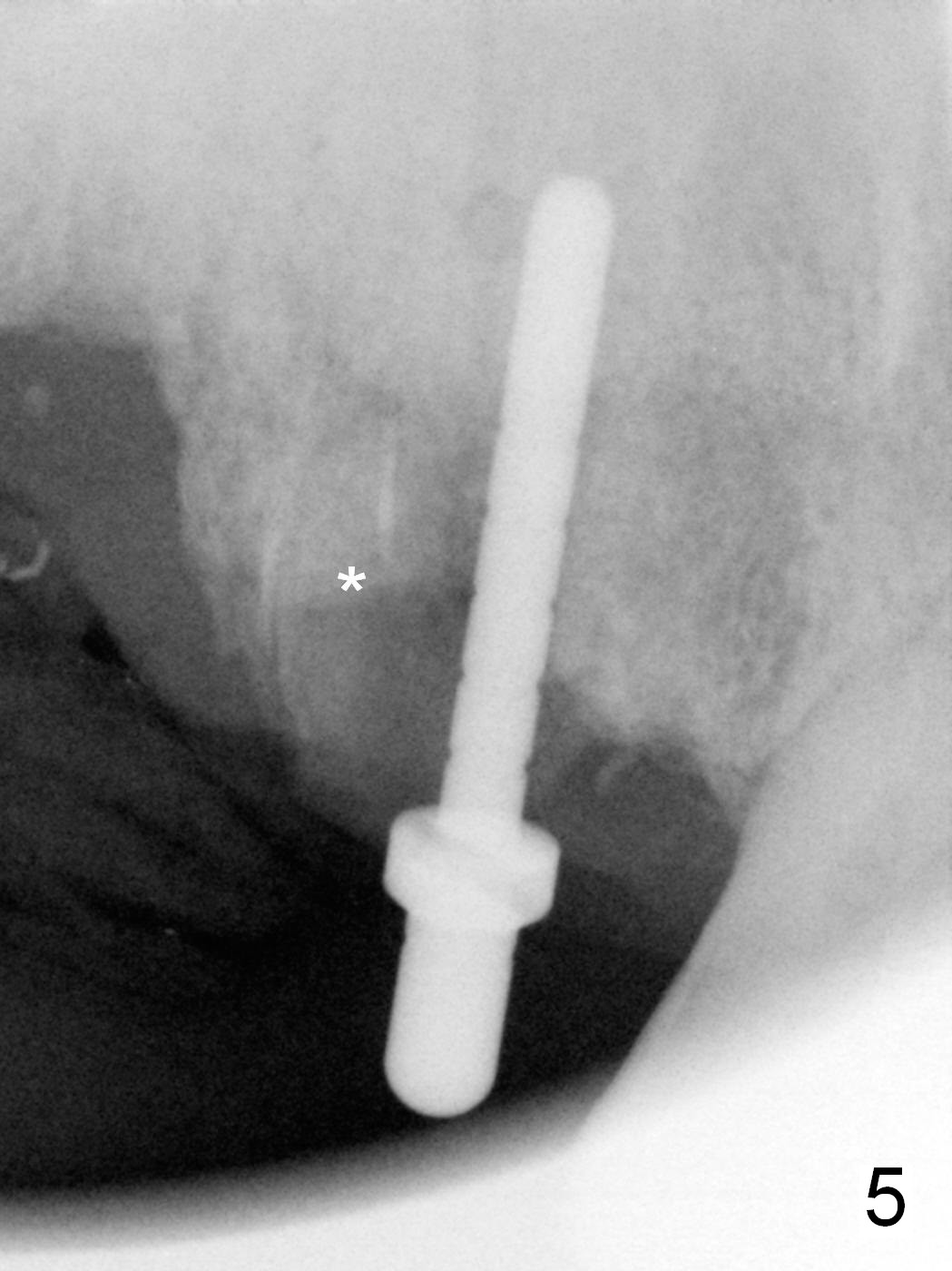
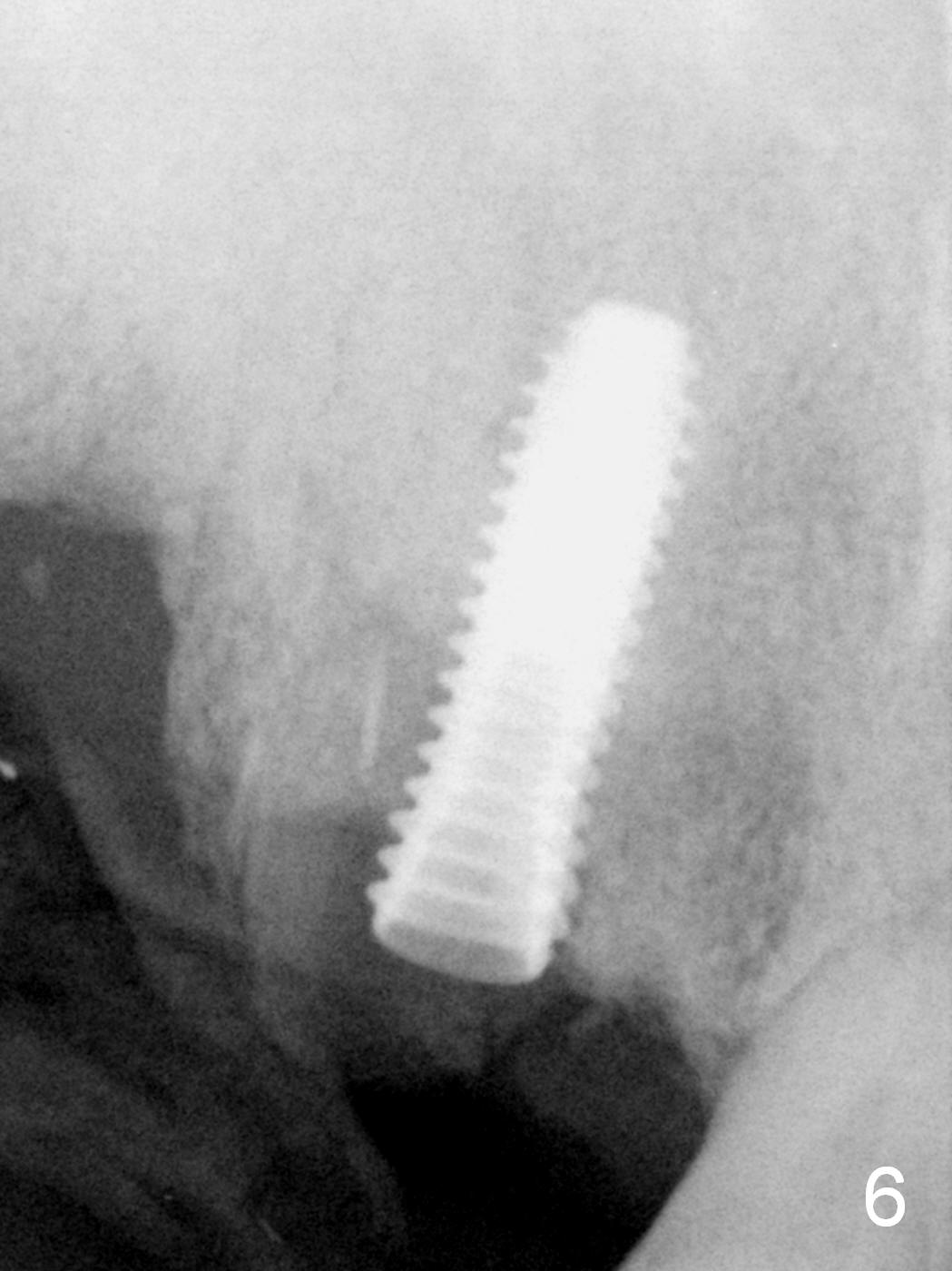
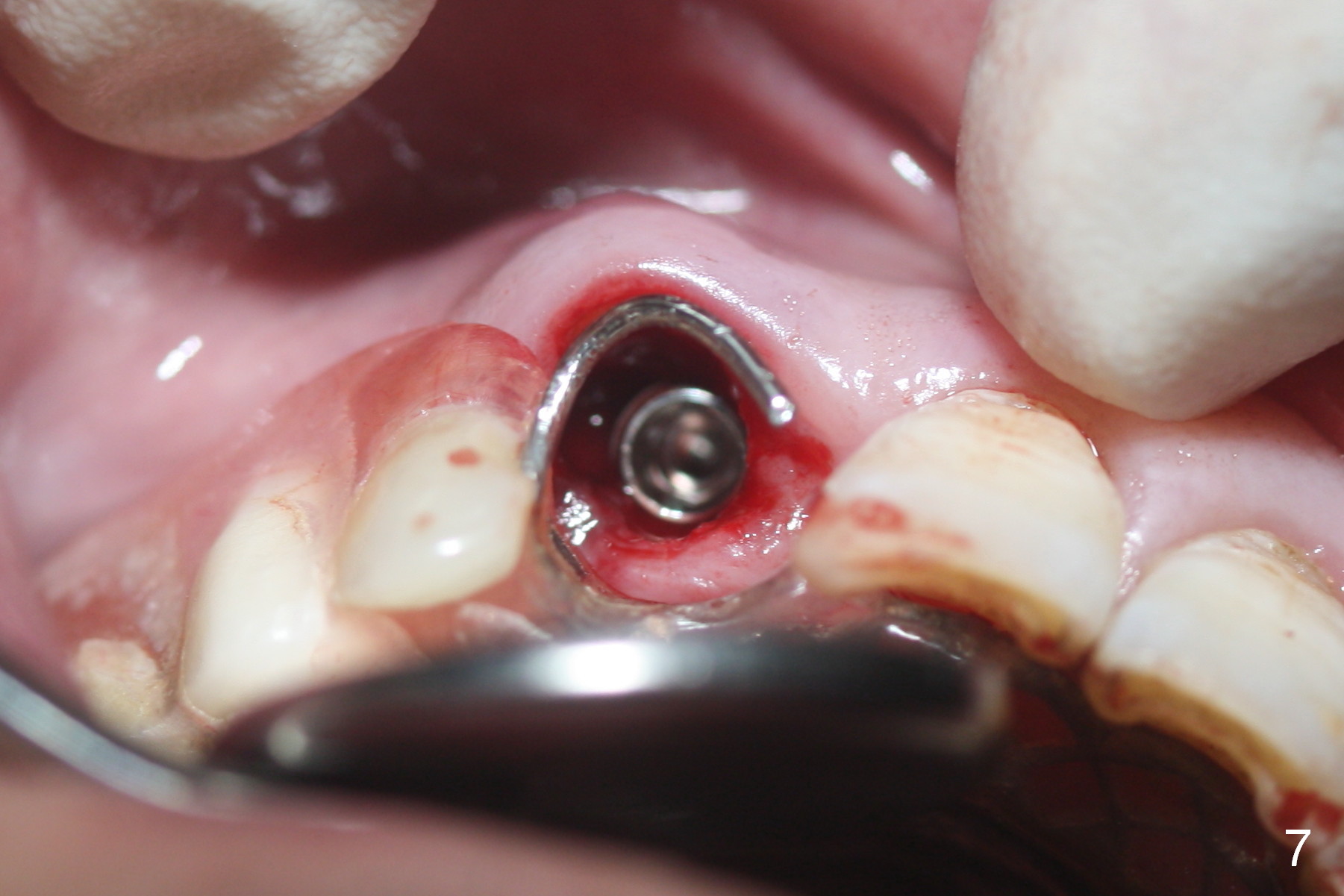
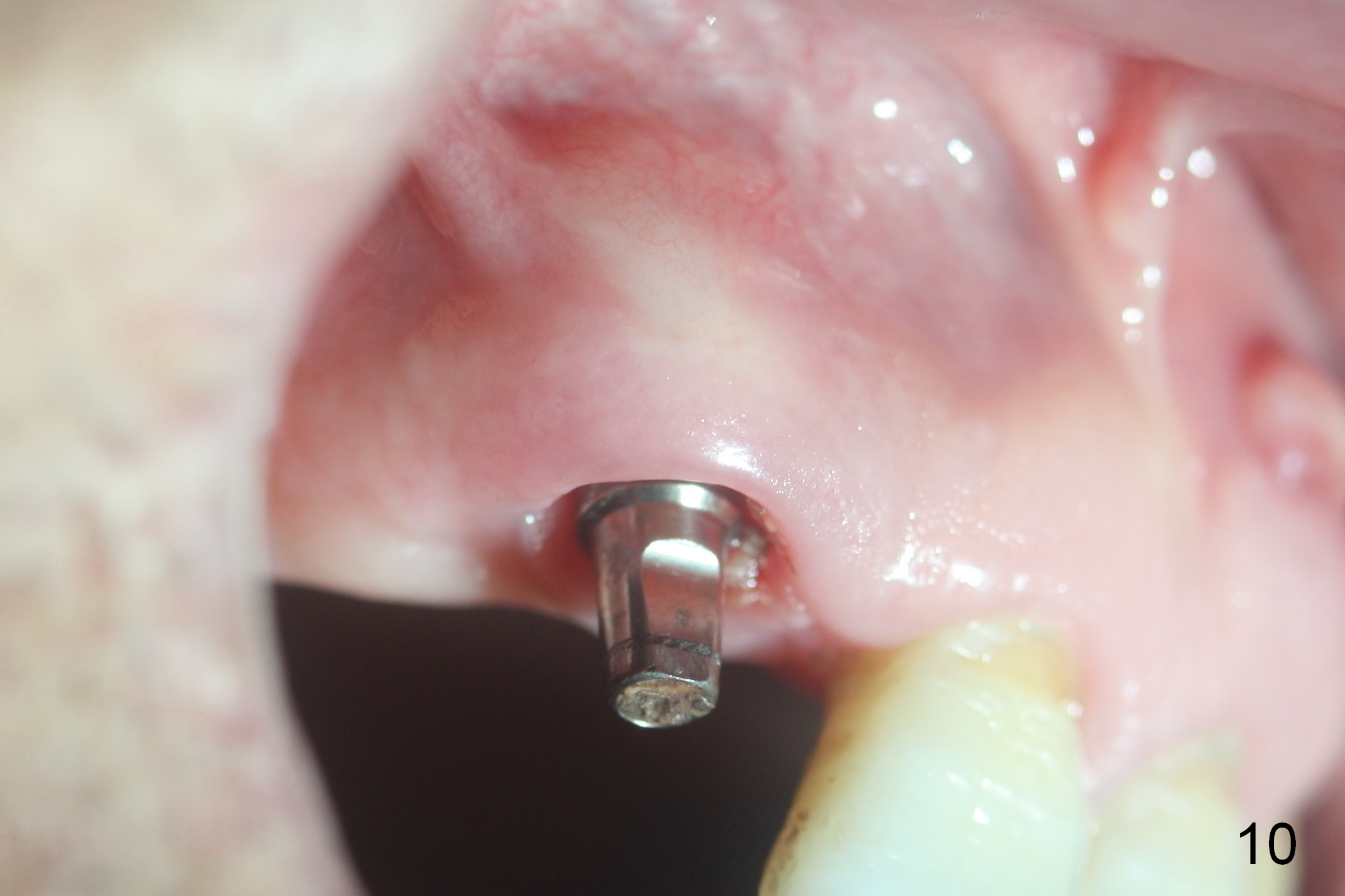
The tooth at #6 was used clinically as a lateral incisor (Fig.1: #6). When the RPD is removed, the buccal gingiva looks healthy (Fig.2). After socket shield is presumably finished (Fig.4 ^), osteotomy is initiated in the palatal aspect of the socket (Fig.3,4). The trajectory is acceptable (Fig.5), but the presence of the apex of the root is ignored (Fig.5 *) until a 4.5x15 mm implant is placed (Fig.6, 50 Ncm). Since there is no apparent periapical radiolucency, the radicular apex is not intended to be removed. The implant remains palatal (Fig.7). A definitive abutment (4.5x5(3) mm) is placed with .5-1 mm bone graft (Fig.8 *, mixed with autogenous bone and Osteogen). An immediate provisional is fabricated to hold the partial and the bone graft in place.
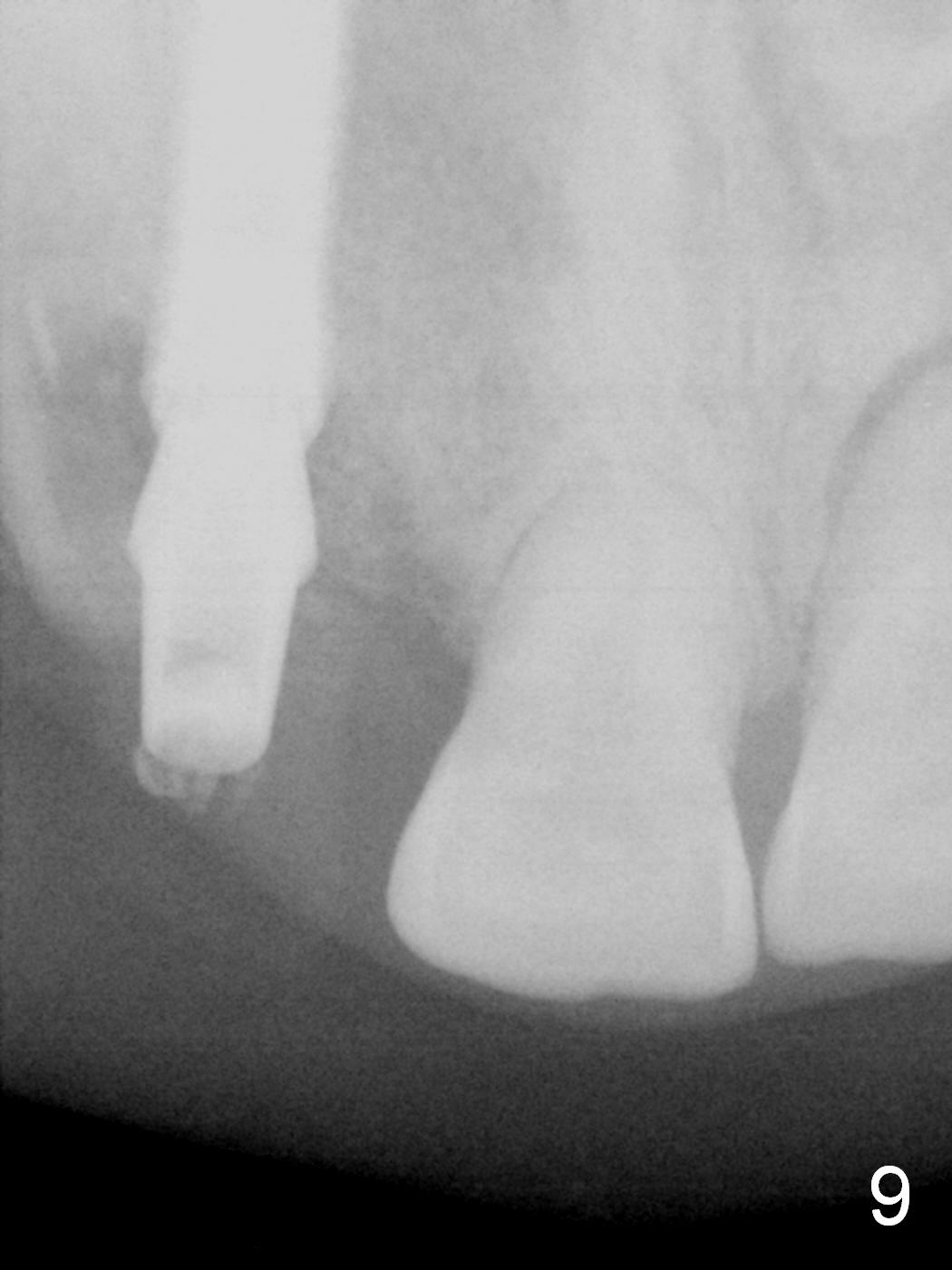
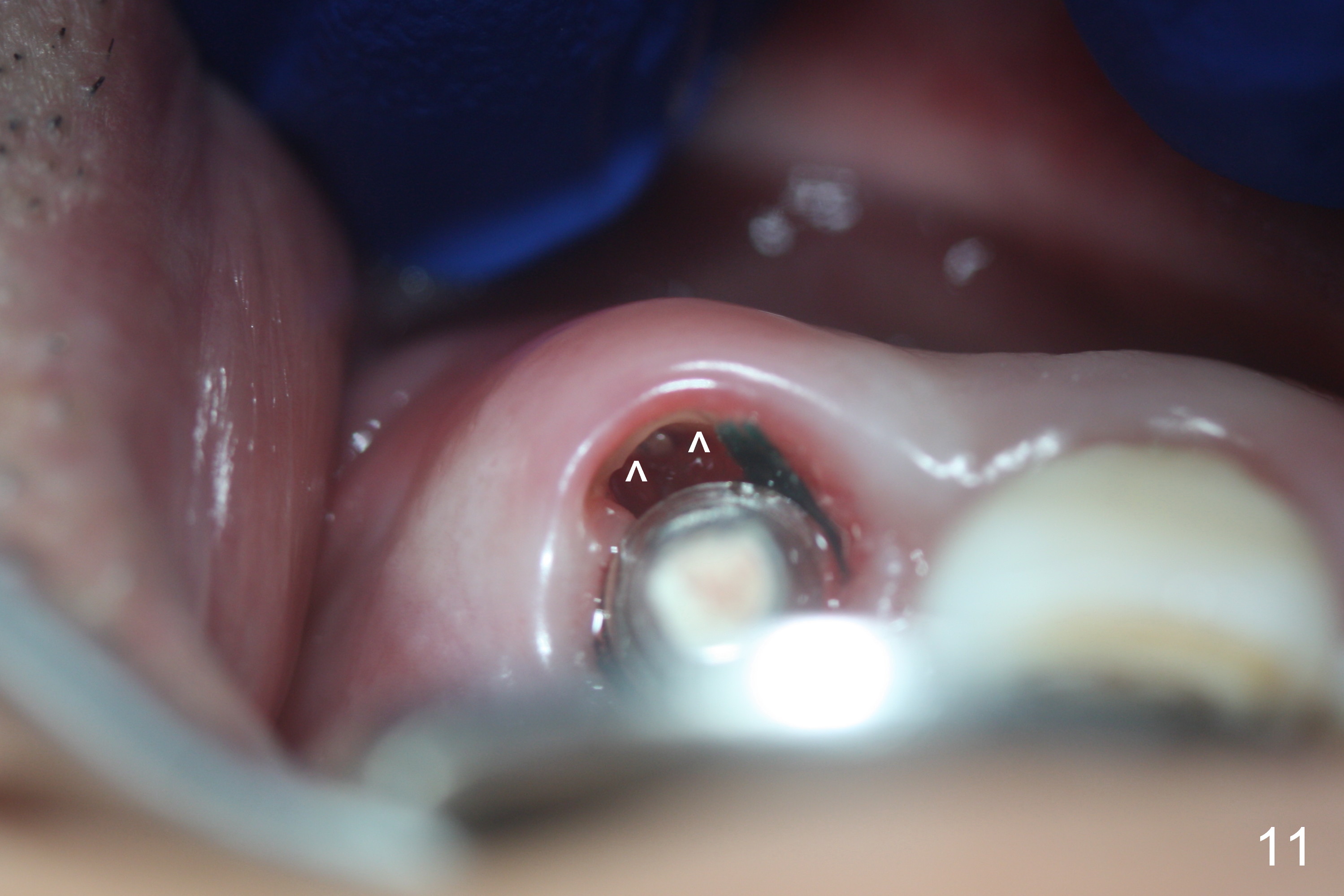
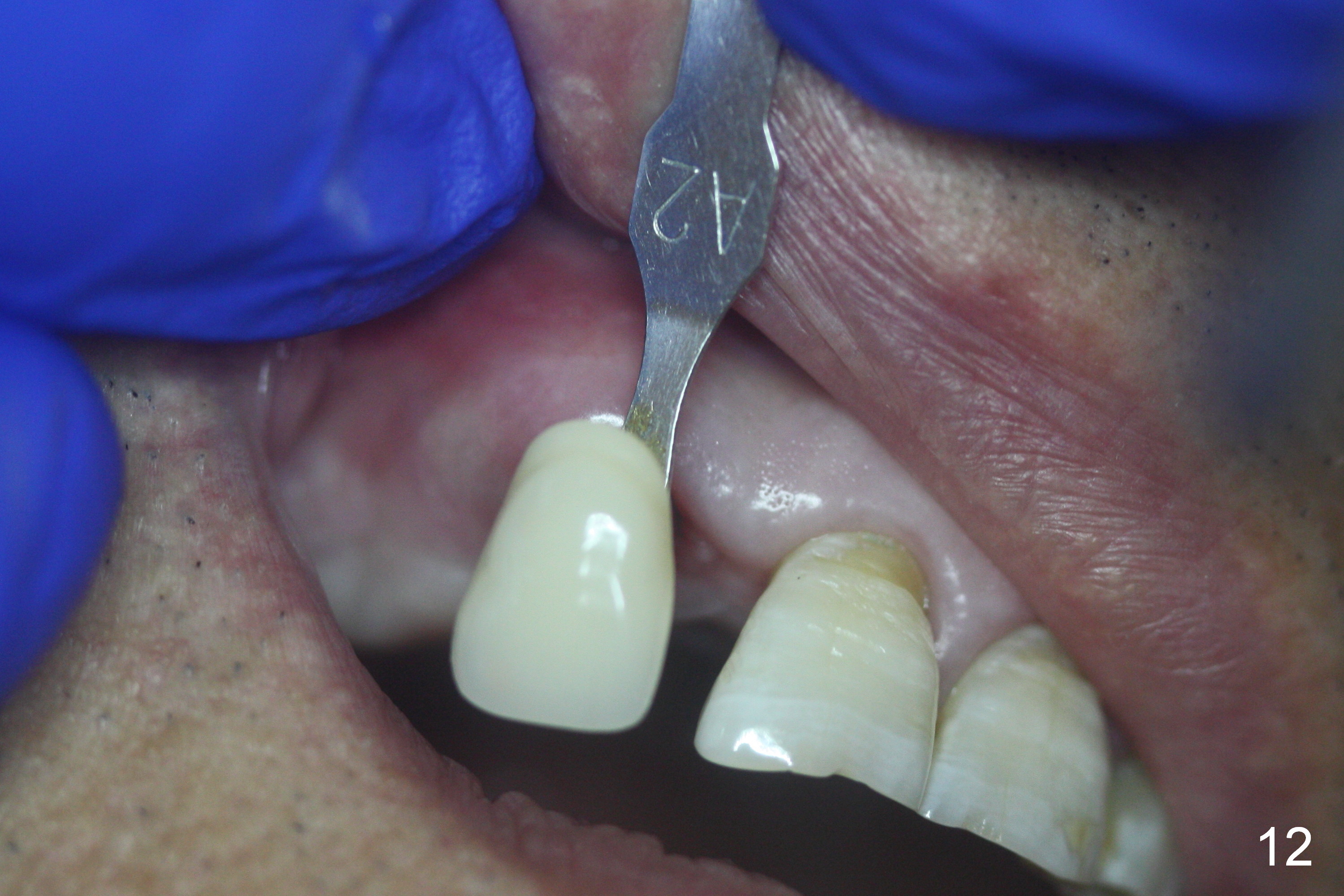
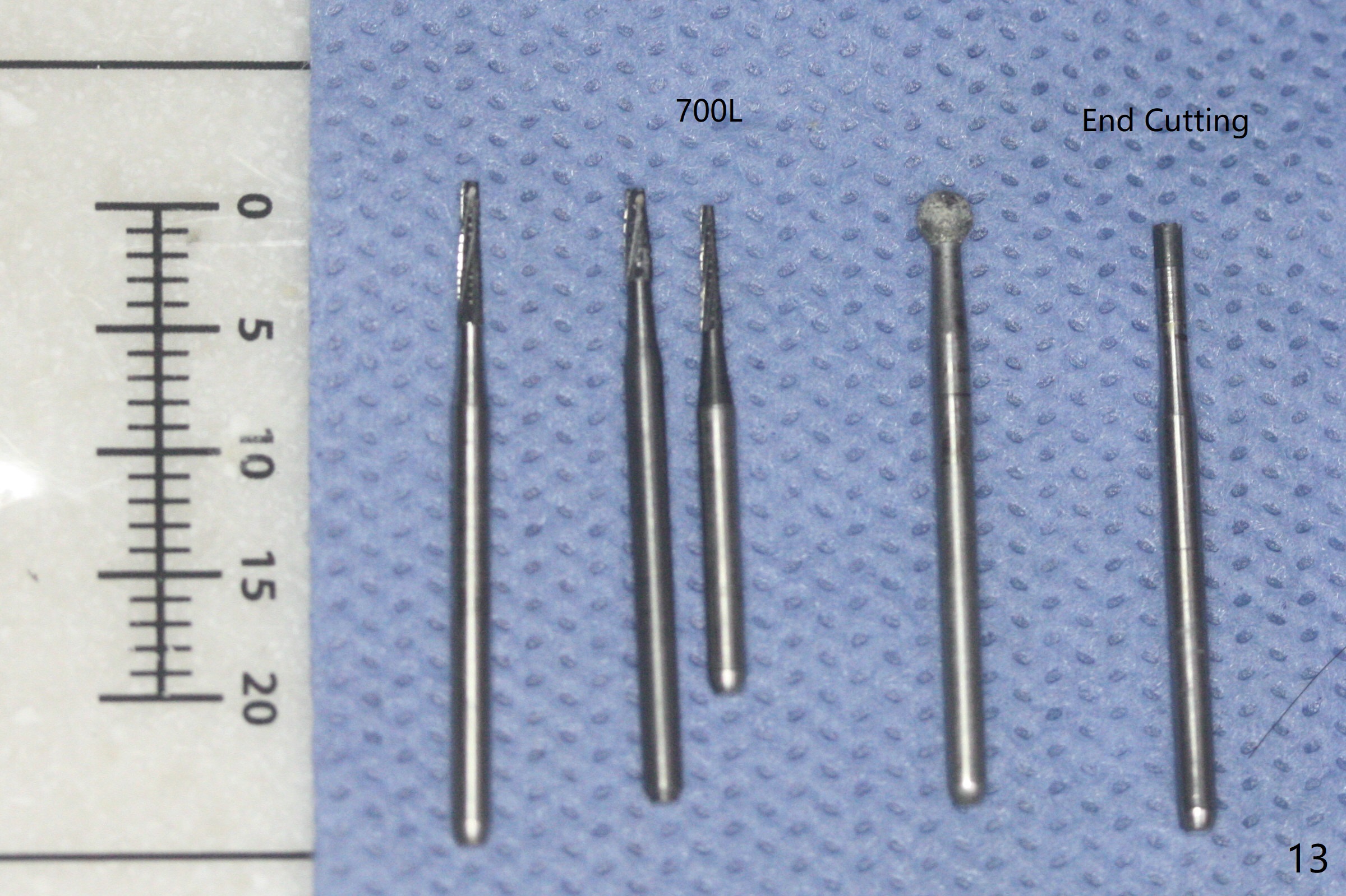
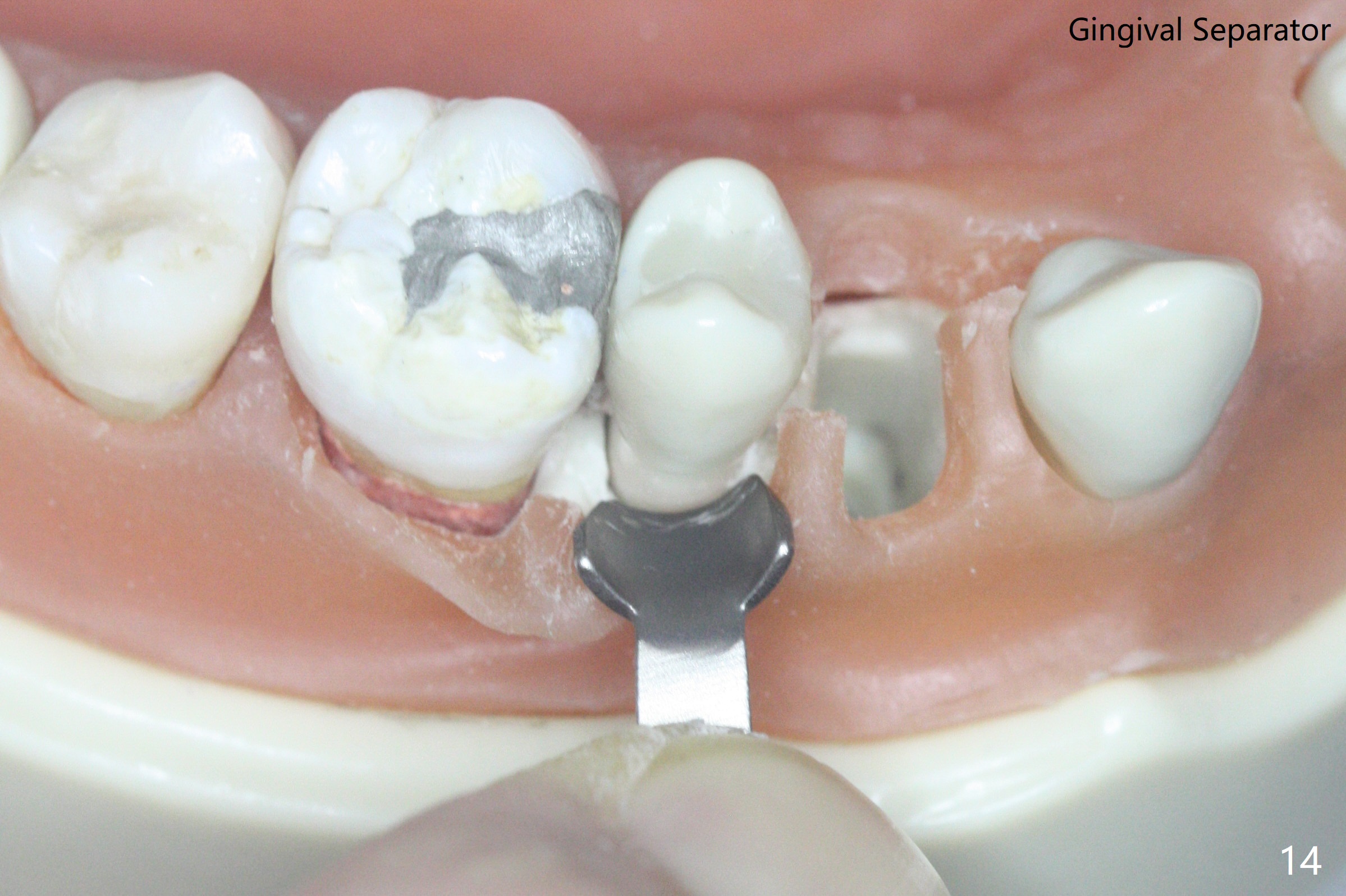
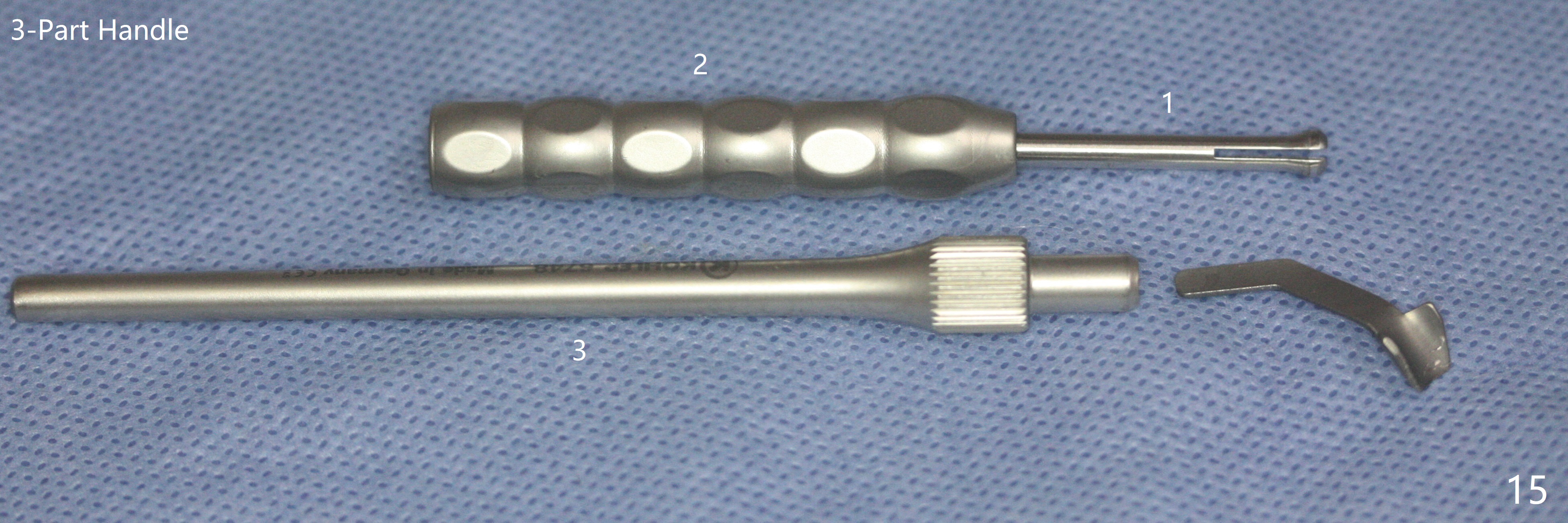
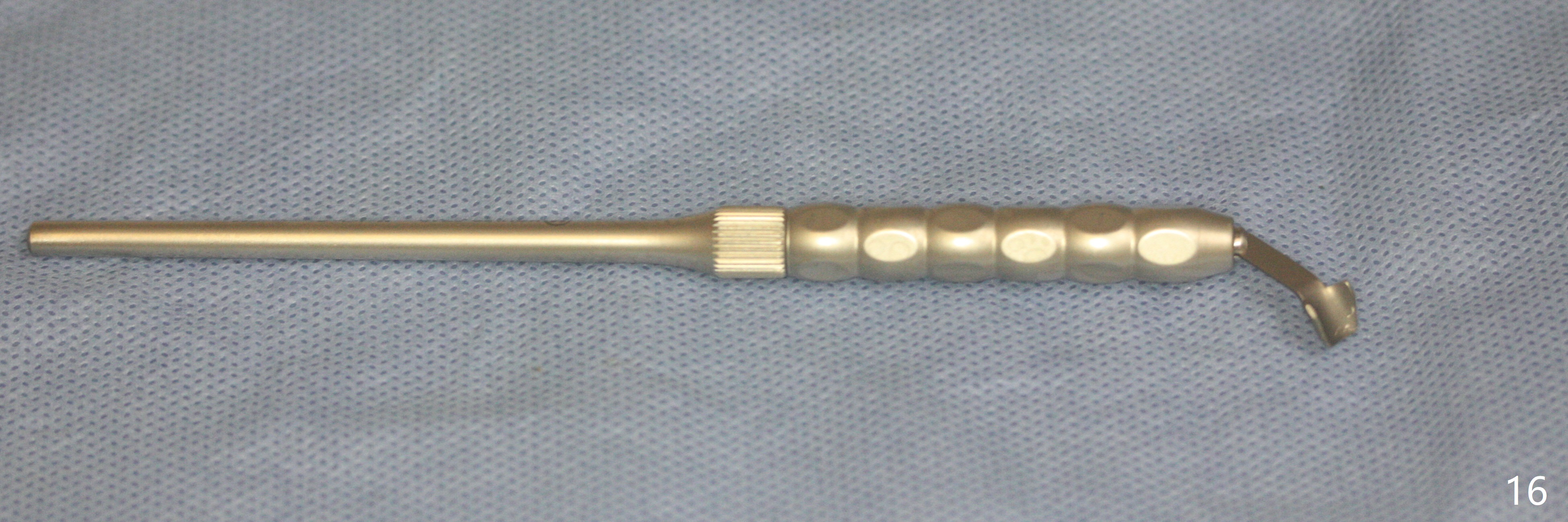
The patient returns for definitive restoration 3.5 months postop (Fig.9-12). The bone graft does not stay between the socket shield (Fig.11 arrowheads) and the implant. Considering the position of the residual root (Fig.1) and clasps (Fig.3,7), make the crown looks like a canine or lateral incisor with a possible mesial diastema. The gingiva looks healthy 14 months post cementation. 盾形成钻头(图十三):外科长度裂钻(左边两个,与第三个修复裂钻相比),最左边25毫米,最好再长些,例如31毫米,用于上尖牙。第四个金刚圆钻用于形成半月形(盾);最后一个End Cutting bur(末端钻)用于缩短盾的长度,而不损伤牙龈。有些公司(例如USTOMED)生产保护牙龈工具(图十四),牵拉牙龈。这个工具成对:左右各一个。这个工具需要三个部分把柄(图十五,十六)固定,有利于口内操作。
Return to
Upper
Canine Immediate Implant
Technicians #18
27
31
Xin Wei, DDS, PhD, MS 1st edition 02/02/2016, last revision 03/24/2021